
Description
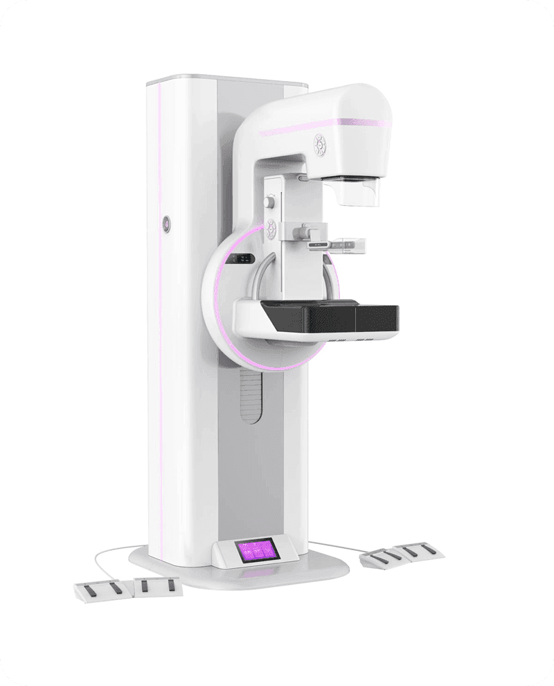
Digital Medical X-ray Radiographic System Multi-functional Diagnostic Dynamic DR Foinoe FN-650MA-2 Remote Table Series

| FN-650MA-2 PACKING LIST | ||||||
| Systems | Parts | Quantity | Model | |||
| Detector | Dynamic Flat Panel Detector | 1 | / | |||
| DXRay Diagnostic Workstation | 1 | / | ||||
| X-Ray System | High Frequency Generator | 1 | / | |||
| Control Cabinet | 1 | / | ||||
| X-Ray Tube | 1 | / | ||||
| High Voltage Cables | 2 | / | ||||
| Collimator | 1 | / | ||||
| Motion Control System | Remote Control Table | 1 | / | |||
| Remote-Control | 1 | / | ||||
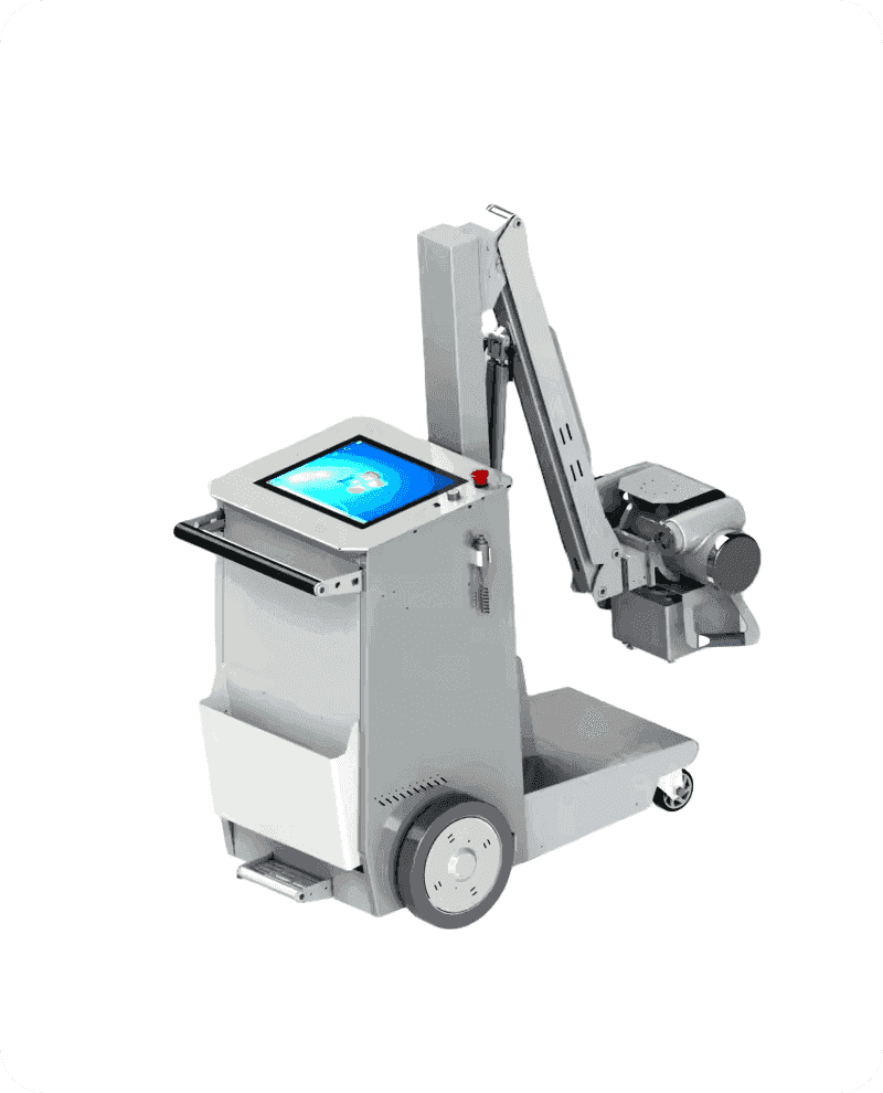
| Mobile Remote Table(Optional) | 1 | / | ||||
| Accessories | Grid | 1 | / | |||
| Report Laser Printer | 1 | / | ||||
| Microphone/Speaker | 1 | / | ||||
| FN-650MA-2 SPECIFICATIONS | |||||
| Part | Remark | ||||
| Dynamic Flat Panel Detector | Dynamic Flat Panel Detector Type | a-Si Flat Panel Detector | |||
| Scintillator Screen Type | Csl (Cesium lodide) | ||||
| Effective View Field | 17 × 17 in. (43 × 43 cm) | ||||
| Static Pixel Matrix | 3072 x 3072 | ||||
| Dynamic Pixel Matrix | 1536 x 1536 | ||||
| Spot-Film Pixel Matrix | 3072 x 3072 | ||||
| Spot-Film Preparation Time | ≤0.8s | ||||
| Output Grayscale | 16bit | ||||
| Preview Image Time | ≤3s | ||||
| Maximum Spatial Resolution | 3.5 lp/mm | ||||
| Detector Movement Range | ≥950mm | ||||
| X-ray Tube | Tube Focus | 0.6mm/1.0mm | |||
| Max Output Voltage | 150kV | ||||
| Heat Capacity | 330kHU | ||||
| Output Power | 30kW/50kW | ||||
| High Frequency Generator | Power Requirement | 380V with 3 Phases Wires | |||
| Maximum Output Power | 50kW | ||||
| Output Voltage for Radiography | 40kV-150kV | ||||
| Output Voltage for Fluoroscopy | 40kV-120kV | ||||
| Output Current for Radiography | 10mA-650mA | ||||
| Output Current for Fluoroscopy | 0.5mA-10mA | ||||
| Range of mAs | 0.4mAs-630mAs | ||||
| Input Power Frequency | 50Hz/60Hz | ||||
| Auto Brightness Selection Function(ABS) | |||||
| Remote Control Table | Table Size | 2100mm x 880mm x 730mm | |||
| Table Lateral Movement | 250mm | ||||
| SID | 1000-1800mm | ||||
| Table Rotate Range | -15° – +90° | ||||
| Tube Column Rotate Range | -35° – +35° | ||||
| High Voltage Cables of X-ray Machine | Length: 8m | ||||
| Rotation Angle of Foot Pedal | 360° | ||||
| One-click in Place Function | Chest Position or Table Position | ||||
| Remote-Control | 1.One-click in Place Function:One-click to Chest Position or Table Position | ||||
| 2.Exposure fluoroscopy switch: Exposure and fluoroscopy can be prohibited | |||||
| to ensure the safety of doctors and prevent accidental exposure | |||||
| Mobile Remote Table (Optional) | Height range | 770mm – 1170mm | |||
| 1.One-click in Place Function:One-click to Chest Position or Table Position | |||||
| 2.Exposure fluoroscopy switch: Exposure and fluoroscopy can be prohibited to ensure the safety of doctors and prevent accidental exposure | |||||
| Grid | Grid Ratio | 10:01 | |||
| Grid Focus | 120 cm | ||||
| Support manual removal | |||||
| Collimator | Power | 150W(24VAC) | |||
| Auto Collimation | Yes | ||||
| Inherent Filtering | ≤1.5mmAl@120KV (XS-D2) | ||||
| Workstation (can be customized) | CPU | INTEL-I5 | |||
| RAM | 4G | ||||
| Hard Disk | 500G | ||||
| Display | 1280*1024 pixels or above | ||||
| CD/DVD recording/burning | |||||
| Software | 1.Patient management: Manual registration, Automatic Query from Worklist | ||||
| 2.Image acquisition: Automatic Window Adjustment, Automatic Sending,Static Image Acquisition, Dynamic Image Acquisition,Video Save, Playback,Bone Stitching(Optional) | |||||
| 3.Image processing: Image Correction, Image Clipping,Automatic Image Segmentation,EAE Image Enhancement Processing , IEQ Image Processing | |||||
| 4.Image viewer: Window/Level Adjustment, Image Flip, Image Rotation, Image Magnification, Restore | |||||
| 5.Medical report: Automatic Loading of Patient Information, Expert Template | |||||
| 6.Film printing: Supports Standard DICOM 3.0 Printer | |||||
| 7.DICOM transmission: Complies with the standard DICOM 3.0 PACS and Workstation | |||||
| 8. Fluoroscopy amplification: 3 step : 17/15,17/12,17/9. | |||||
| 9. Anatomically programmed radiography (APR) : auto pre-set parameter link to the chosen body part | |||||
| 10. Automatical collimator: After choose the body part, collimator can adjust the suitable window size, changing SID could aslo bring automatical adjustment of collimator | |||||


Reviews
There are no reviews yet.